Laboratory of
Experimental Myopia
University Eye Hospital
Dept. of Pathophysiology of Vision and Neuroophthlmology
Division of Experimental Ophthalmology
Röntgenweg 11, D-72076 Tübingen, Germany
PD Dr.rer.nat. Frank Schaeffel
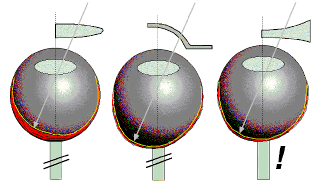
| Placing a positive lens in front of the eye moves the image plane in front of the retina. In response, the choroid (red) behind the retina (green) thickens and moves the retina towards the image plane. Subsequently, the axial growth of the eye stops until the focal length of cornea and lens have sufficiently increased. The mechanism operates locally, even without connection to the brain. The mechanism does not depend on retinal dopamine. | Covering the eye with a frosted, translucent occluder which reduces image contrast causes myopia selectively in the "deprived" areas ("deprivation myopia"). Neither an optic nerve nor ganglion cells are necessary. The reduction in retinal image contrast causes a decline in dopamine release from the retina. Myopia can be prevented by drugs that interact with dopamine release. |
Placing a negative lens in front of the eye moves
the image plane behind the retina. To catch the image, the eye grows more,
selectively in the defocused areas. However, the growth is reduced without
optic nerve. During compensation of the imposed refractive error, the retinal
dopamine release is reduced. The compensation can be suppressed by drugs
which enhance dopamine release or interact with dopamine receptors. |
| Co-workers and projects: |
|
||

Dr. Marita Feldkaemper |

Dipl.Biol Hartmut Schwahn |

Dipl.Biol Sibylle Ohngemach |
Dipl.Biol Sigrid Diether |
PTA Gabi Hagel |

cand.med. Florian Gekeler |

cand.med. Hakan Kaymak |
cand.med. Silke Thomas |
Homepage of the University Tübingen
Homepage of the University
Eye Hospital Tübingen
